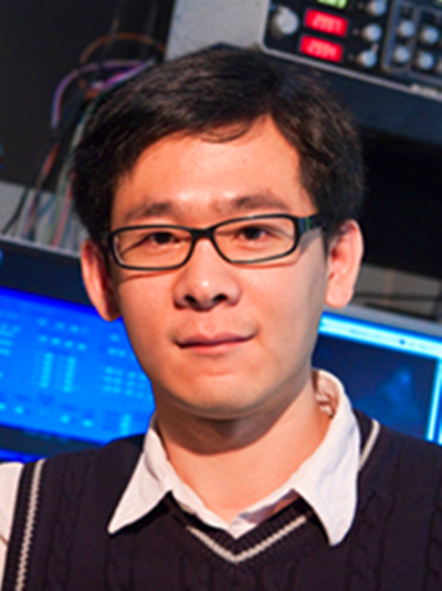
許迎科,1982年出生於浙江東陽。1999年至2008年就讀於浙江大學,先後獲本科及博士學位。2008年至2012年,於美國耶魯大學醫學院從事科學研究工作,先後擔任博士後及研究助理科學家。2012年底回國任教至今,先後獲浙江大學求是青年學者,浙江省傑出青年基金,入選浙江省151人才工程,主要從事生物醫學光子學與細胞生物學的套用研究工作。作為項目負責人主持國家自然科學基金項目4項;參與科技部973項目、國家重點研發計畫與基金委重大儀器專項等多項縱向研究課題。目前擔任美國耶魯大學兼職教授(Adjunct Professor)、Journal of Cellular and Molecular Medicine雜誌副主編、Engineering雜誌青年通訊專家等。
基本介紹
- 中文名:許迎科
- 畢業院校:浙江大學
- 學位/學歷:博士
- 專業方向:生物光子學、生物物理、光遺傳學、活細胞顯微成像、定量圖像處理
- 職務:院長助理
學術兼職,獎勵榮譽,發表論文,
學術兼職
紐約科學院(NYAS),美國糖尿病學會(ADA),美國細胞生物學學會(ASCB),中國細胞生物學學會(CSCB),中國生物物理學會(BSC),中國生物醫學工程學會等學術組織 會員
-美國耶魯大學醫學院 兼職教授(Adjunct Professor)
-浙江大學醫學院附屬邵逸夫醫院 兼聘教授
-浙江省光學學會 理事 (Fellow)
-Journal of Cellular and Molecular Medicine (SCI; IF5=5.02) Associate Editor
-Engineering (SCI; IF=4.6) Corresponding Expert
獎勵榮譽
2011 美國細胞生物學會Travel Award (ASCB Travel Award)
2012 第三屆國際膜生物學會議Biochemical Journal Best Poster Award
2012 浙江大學求是青年學者
2013 浙江大學優秀青年教師計畫-“紫金計畫”
2013 浙江大學求是學院 優秀班主任
2014 浙江大學校級 優秀班主任
2015浙江大學青年教師教學技能競賽優勝獎
2016 浙江大學信息學部青年創新獎(僅4人入選)
2016 浙江大學金秀獎教金獲得者
2016 浙江大學生儀學院先進工作者
2017浙江省傑出青年基金獲得者
2017浙江大學校級先進工作者
2018浙江大學生儀學院先進工作者
2018入選浙江省151人才工程
發表論文
Chen Y, Liu W, Zhang Z, Zheng C, Huang Y, Cao R, Zhu D, Xu L, Zhang M, Zhang YH, Fan J, Jin L, Xu Y, Kuang C*, Liu X*. Multi-color live-cell super-resolution volume imaging with multi-angle interference microscopy. Nature Communications 2018, 9: 4818.
- Zheng C, Zhao G, Liu W, Chen Y, Zhang Z, Jin L,Xu Y, Kuang C, Liu X*. Three-dimensional super-resolved live cell imaging through polarized multi-angle TIRF.Optics Letters2018, 43:1423-1426.
- Qi Y, Chen J, Liu X, Zhou X, Fan J, Shentu P, Idevall-Hagren O,Xu Y*. Development of a wireless controlled LED array for tunable optogenetic control of cellular activities.Engineering2018, 4: 745-747.
- Zhou X, Wang J, Chen J, Qi Y, Nan D, Jin L, Qian X, Wang X, Chen Q*, Liu X,Xu Y*.Optogenetic control of epithelial-mesenchymal transition in cancer cells.Scientific Reports2018, 8:14098.
- Cao R, Chen Y, Liu W, Zhu D, Kuang C*,Xu Y*,Liu X. Inverse matrix based phase estimation algorithm for structured illumination microscopy.Biomedical Optics Express2018, 9:5037-5051.
- Wang W, Zhao G, Kuang C*, Xu L, Liu S, Sun S, Shentu P, Yang Y,Xu Y,Liu X*. Integrated dual-color stimulated emission depletion (STED) microscopy and fluorescence emission difference (FED) microscopy.Optics Communications2018, 423:167-174.
- Xiu P, Liu Q, Zhou X,Xu Y*,Kuang C, Liu X. Analogous on-axis interference topographic phase microscopy (AOITPM).Journal of Microscopy2018, 270: 235-243.
- Jin L, Zhou X, Xiu P, Luo W, Huang Y, Yu F, Kuang C, Sun Y, Liu X, Xu Y*. (2017) Imaging and reconstruction of cell cortex structures near the cell surface. Optics Communications 402, 699-705.
- Xu Y, Toomre D, Bogan JS, Hao M*. Excess cholesterol inhibits glucose-stimulated fusion pore dynamics in insulin exocytosis.Journal of Cellular and Molecular Medicine2017, 21:2950-2962.
- Zhang D, Wang R, Xiang Y Kuai Y, Kuang C, Badugu R,Xu Y, Wang P, Ming H, Liu X, Lakowicz JR. Silver nanowires for reconfigurable bloch surface waves.ACS Nano2017, 11:10446-10451.
- Li H, Mao Y, Yin Z*, Xu Y*. (2017) A hierarchical convolutional neural network for vesicle fusion event classification. Computerized Medical Imaging and Graphics 60, 22-34.
- Huang Y, Zhu D, Jin L, Kuang C, Xu Y*, Liu X. (2017) Laser scanning saturated structured illumination microscopy based on phase modulation. Optics Communications 396, 262-266.
- Fan J, Zhou X, Wang Y, Kuang C, Sun Y, Liu X, Toomre D, Xu Y*. (2017) Differential requirement for N-ethylmaleimide-sensitive factor in endosomal trafficking of transferrin receptor from anterograde trafficking of vesicular stomatitis virus glycoprotein G. FEBS Letters 591, 273-281. (Editor’s Highlights)
- Li H, Ou L, Fan J, Xiao M, Kuang C, Liu X, Sun Y, Xu Y*. (2017) Rab8A regulates insulin-stimulated GLUT4 trafficking in C2C12 myoblasts. FEBS Letters 591, 491-499. (Editor’s Choice)
- Zhou X, Shentu P, Xu Y*. (2017) Spatiotemporal regulators for insulin-stimulated GLUT4 vesicle exocytosis. Journal of Diabetes Research. ID 1683678. doi:10.1155/2017/1683678.
- Jin L, Wu J, Xiu P, Fan J, Hu M, Kuang C, Xu Y*, Zheng X, Liu X. (2017) High-resolution 3-D reconstruction of microtubule structures by quantitative multi-angle total internal reflection fluorescence microscopy. Optics Communications 395, 16-23.
- Liu X, Kuang C, Hao X, Pang C, Xu P, Li H, Liu Y, Yu C, Xu Y, Nan D, Shen W, Fang Y, He L, Liu X*, Yang Q*. (2017) Fluorescent nanowire ring illumination for wide-field far-field subdiffraction imaging. Physical Review Letters 118, 076101.
- Kuang C, Ma Y, Zhou R, Zheng G, Fang Y, Xu Y, Liu X*, So PTC*. (2016) Virtual k-space modulation optical microscopy. Physical Review Letters 117, 028102.
- Xu Y*, Nan D, Fan J, Bogan JS, Toomre D*. (2016) Optogenetic activation reveals distinct roles of PI(3,4,5)P3 and Akt in adipocyte insulin action. Journal of Cell Science. 129, 2085-2095.
- Chai GH, Xu Y, Chen SQ, Cheng B, Hu FQ, You J, Du YZ, Yuan H*. (2016) Transport mechanisms of solid lipid nanoparticles across Caco-2 cell monolayers and their related cytotoxicology. ACS Appl. Mater. Interfaces. 8(9): 5929-5940.
- Wang Y, Ma Y, Kuang C*, Fang Y, Xu Y, Liu X, Ding Z. (2015) Dual-mode super-resolution imaging with stimulated emission depletion microscopy and fluorescence emission difference microscopy. Appl. Opt. 54(17), 5425-5431.
- Wu J, Xu Y, Feng Z, Zheng X*. (2015) Automatically identifying fusion events between GLUT4 storage vesicles and the plasma membrane in TIRF microscopy image sequences. Comput. Math. Methods Med. 2015;2015:610482. doi: 10.1155/2015/610482.
- Cai H, Wang Y, Kuang C, Ge J, Xu Y, Liu X. (2015) Sub-diffraction imaging with total internal reflection fluorescence (TIRF) microscopy by stochastic photobleaching. Journal of Modern Optics312, 62-67.
- Ma Y, Kuang C, Fang Y, Ge B, Mao X, Shen S, Xu Y, Liu X. (2015) Particle localization with total internal reflection illumination and differential detection. Optics Communications 339, 14-21.
- Rong Z, Li S, Kuang C, Xu Y, Liu X. (2014) Real-time super-resolution imaging by high-speed fluorescence emission difference microscopy. Journal of Modern Optics 61, 1364-1371.
- Wang Y, Kuang C, Li S, Hao X, Xu Y, Liu X. (2014) A 3D Aligning method for stimulated emission depletion microscopy using fluorescence lifetime distribution. Microscopy Research and Technique 77, 935-940.
- Xiu P, Zhou X, Kuang C, Xu Y, Liu X. (2014) Controllable tomography phase microscopy. Optics and Lasers in Engineering 66, 301-306.
- Chenouard N, Smal I, Chaumont F, et al. Liang L, Duncan J, Shen H, Xu Y, et al. Olivo-Marin J, Meijering E. (2014) Objective comparison of particle tracking methods. Nature Methods 11, 281-290. (IF=23.57) (封面文章)
- Zhang Y, Tang W, Zhang H, Niu X,Xu Y, Zhang J, Gao K, Pan W, Boggon TJ, Toomre D, Min W, Wu D. (2013) A network of interactions enables CCM3 and STK24 to coordinate UNC13D-driven vesicle exocytosis in neutrophils.Developmental Cell27, 215-226. (IF=12.86)
- Wang Y, Kuang C, Gu Z,Xu Y, Li S, Hao X, Liu X. (2013) Time-gated stimulated emission depletion nanoscopy.Optical Engineering52, 093107-093107.
- Bogan JS, Xu Y, Hao M. (2012) Cholesterol accumulation increases insulin granule size and impairs membrane trafficking. Traffic 11, 1466-1480. (IF=4.92)
- Xu Y, Melia TJ, Toomre DK. (2011) Using light to see and control membrane traffic. Current Opinion in Chemical Biology 15, 822-830. (IF=9.85)
- Xu Y, Rubin BR, Orme CM, Karpikov A, Yu C, Bogan JS, Toomre DK (2011) Dual-mode of insulin action controls GLUT4 vesicle exocytosis. Journal of Cell Biology 193, 643-653. (IF=10.26)
- Feng L,Xu Y, Yang Y, Zheng X (2011) Multiple dense particle tracking in fluorescence microscopy images based on multidimensional assignment. Journal of Structural Biology173, 219-228. (IF=3.5)
- Chen Q, Lu G, Wang Y, Xu Y, Zheng Y, Yan L, Jiang Z, Yang L, Zhan J, Wu Y, Zhou J. (2009) Cytoskeleton disorganization during apoptosis induced by curcumin in A549 lung adenocarcinoma cells. Planta Medica 75, 808-813.
- Deng N, Xu Y, Sun DY, Hua PF, Zheng XX, Duan HL (2009) Image processing for fusion identification between the GLUT4 storage vesicles and plasma membrane. Journal of Signal Processing Systems54, 115-125.
- Wu X, Li J, Xu Y, Xu K, Zheng X (2008) Three-Dimensional Tracking of GLUT4 Vesicles in TIRF Microscopy. Journal of Zhejiang University- Science A 9(2), 232-240.
- Xu Y, Xu K, Li J, Feng L, Zheng X (2007) Bi-directional transport of GLUT4 vesicles near the plasma membrane of primary rat adipocytes. Biochemical and Biophysical Research Communications 359(1), 121-128.
- Xu Y, Li J, Xu K, Zheng X (2007) Real-time and dynamic tracking of single mobile GLUT4 vesicles in primary rat adipocytes: application of total internal reflection fluorescence microscopy. Journal of Zhejiang University-Engineering 41(12), 2112-2116. (EI)

