編輯推薦
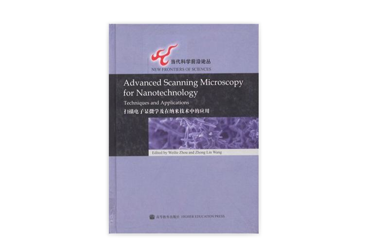
《掃描電子顯微學及在納米技術中的套用》的特點是各個章節都是由本領域裡世界級資深的研究組所撰寫。其他貢獻者則是來自這些技術和設備的國際知名供應商中的技術套用專家。《掃描電子顯微學及在納米技術中的套用》不僅可供納米材料研究者使用,同時也是一本面向高校師生和掃描電子顯微鏡工作者的教材和參考書。
內容簡介
在過去的10年中,納米技術的飛速發展使得掃描電子顯微鏡成為了一種分析和構建新納米材料、結構和器件不可缺少的有力的工具。新納米材料的發現需要通過先進的分析技術和技能來獲取高質量的圖片,從而幫助我們理解納米結構,以達到改進合成方法和提高性能的目的。例如場發射槍、背散射電子的探測、X射線元素的圖像化等,已經很大程度地提高了掃描電子顯微鏡在納米材料分析中的套用。除了分析功能之外,掃描電子顯微鏡可以與新發展起來的測控技術相結合,實行原位納米器件的加工、製造和性能表征。這些技術包括納米材料的操控、電子刻蝕、聚焦離子束微加工等。雖然這些技術仍在發展之中,但它們已開始廣泛套用於納米研究的各個領域。
作者簡介
周維列,博士,1993年畢業於中國科學院物理研究所。1994一1997年先後在日本精密陶瓷材料研究所(JFCC)和美國凱斯西部保留大學(CWRU)從事電子顯微學博士後研究。1998年至今任美國紐奧良大學(LJNO)先端材料研究所(AMRI)電鏡室主任。從事電子顯微鏡在材料中的套用近20年,獲美國自然科學基金(NSF)和美國國防部先端研究項目(DARPA)等多項資助。現為美國電子顯微學會和材料學會會員。
王中林博士,美國喬治亞理工學院(Georgia Institute of TechnoIogy)終身教授、校董事講席教授(Regents’Professor)、納米結構表征和器件製造中心主任,中國國家納米科學中心海外主任,美國物理學會資深會員(Fellow)。榮獲美國顯微鏡學會1999年巴頓獎章,喬治亞理工學院2000年和2005年傑出研究獎,2005年Sigma Xi學會持續研究獎,2001年S.T.Li獎金(美國化學學會),美國自然科學基金會CAREER基金。他是1992——2002年10年中納米科技論文引用次數世界個人排名前25位作者之一,其學術論文已被引用10000次以上。
目錄
Chapter 1 Fundamentals of Scanning Electron Microscopy
1.1 Introduction
1.2 Configuration of scanning electron microscopes
1.3 Specimen preparation
1.4 Conclusion
References
Chapter 2 Electron Backscatter Diffraction(EBSD)Technique and Materials Characterization Examples
2.1 Introduction
2.2 Data measurement
2.3 Data analysis
2.4 Applications
2.5 Current limitations and fur
2.6 Conclusion
References
Chapter 3 X-ray Microanalysis in Nanomaterials
3.1 Introduction
3.2 Monte Carlo modeling of nanomaterials
3.3 Case studies
3.4 Conclusion
References
Chapter 4 Low kV Scanning Electron Microscopy
4.1 IntrOductlon
4.2 Electron generation and accelerating voltage
4.3 Why use low kV?
4.4 Using low kV
4.5 Conclusion
References
Chapter 5 E-beam Nanolithography Integrated with Scanning Electron Microscope
5.1 Introduction
5.2 Materials and processing preparation
5.3 Pattern generation
5.4 Pattern processing
5.5 Applications
5.6 Conclusion
References
Chapter 6 Scanning Transmission Electron Microscopy for Nanostructure Characterization
6.1 Introduction
6.2 Imaging in the STEM
6.3 Spectroscopic imaging
6.4 Three-dimensional imaging
6.5 Recent applications to nanostructure characterization
6.6 Future directions
References
Chapter 7 Introduction to ln-situ Nanomanipulation for Nanomaterials Engineering
7.1 Introduction
7.2 SEM Contamination
7.3 Types of nanomanipulators
7.4 End effectors
7.5 Applications of nanomanipulators
7.6 Conclusion
References
Chapter 8 Applications of FIB and DualBeam for Nanofabrication
8.1 Introductlon
8.2 Onboard digital patterning with the ion beam
8.3 FIB milling or CVD deposition with bitmap files
8.4 Onboard digital patterning with the electron beam
8.5 Automation for nanometer control
8.6 Direct fabrication of nanoscale structures
8.7 Conclusion
References
Chapter 9 Nanowires and Carbon Nanotubes
9.1 Introduction
9.2 Ⅲ-V compound semiconductors nanowires
9.3 Ⅱ-VI compound semiconductors nanowires
9.4 Elemental nanowires
9.5 Carbon nanotubes
9.6 Conclusion
References
Chapter 10 Photonic Crystals and Devices
10.1 Introduction
10.2 SEM imaging of photonic crystals
10.3 Fabrication of photonic crystals in SEM
10.4 Conclusion
References
Chapter 11 Nanoparticles and Colloidal Self-assembly
11.1 Introduction
11.2 Metallic nanoparticles
11.3 Mesoporous and nanoporous metal nanostructures
11.4 Nanocrvstalline oxides
11.5 Nanostructured semiconductor and thermoelectric materials
11.6 Conclusion
References
Chapter 12 Nano-building Blocks Fabricated through Templates
12.1 Introduction
12.2 Materials and methods
12.3 Nano-building blocks
12.4 Conclusion
References
Chapter 13 One-dimensional Wurtzite Semiconducting Nanostructures
13.1 Introduction
13.2 Synthesis and fabrication of one—dimensional nanostructures
13.3 One-dimensional metal oxide nanostructures
13.4 Growth mechanisms
13.5 Conclusion
References
Chapter 14 Bio-inspired Nanomaterials
14.1 Introduction
14.2 Nanofibers
14.3 Nanoparticles
14.4 Surface modification
14.5 Conclusion
References
Chapter 15 Cryo-Temperature Stages in Nanostructural Research
15.1 Introduction
15.2 Terminology used in cryo-HRSEM of aqueous systems
15.3 Liquid water,ice,and vitrified water
15.4 History of 10W temperature SEM
15.5 Instrumentation and methods
References

