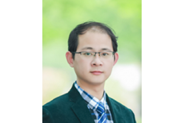
個人經歷
教育背景
2005年 9月 保送浙江大學信息與電子工程學系研究生,導師顧偉康教授
2010年12月 博士畢業於新加坡國立大學(NUS)綜合科學與工程研究生院(NGS),導師為共聚焦顯微鏡的奠基者之一 Colin J. R. Sheppard 教授。
工作經歷
2011年1月至2011年11月:
獲新加坡-美國麻省理工學院(MIT)研究聯盟(SMART)資助,在NUS和MIT,博士後;
2011年11月起2013年:
“諾貝爾獎聖地”美國霍華德休斯醫學研究所(Howard Hughes Medical Institute),助理研究員;
2013年12月起:
浙江大學光電科學與工程學院,教授;
浙江大學醫學院基礎系神經科學研究所,教授;
浙江大學神經生物影像實驗室,主任。
2016年2月起2018年12月:
浙江大學腦科學研究科技聯盟(腦聯盟),秘書長。
2016年2月起至今
浙江大學光電科學與工程學院雷射生物醫學研究所,書記、副所長;
2018年1月至2019年12月:
浙江大學醫學院神經科學研究中心,副執行主任。
2018年7月起至今:
醫學部醫學神經生物學重點實驗室,副主任。
2018年9月起至今:
2018年10月起至今:
科技部光電技術國際聯合研究中心,副主任
2019年3月至2020年3月:
國家腦與腦機融合前沿科學中心(相當於3-4個
國家重點實驗室題量),主任助理
2019年6月起至今:
浙大安德醫學人工智慧研究中心,執行主任
2019年11月起至今:
浙江大學腦科學與腦醫學學院,副院長(副處級)
2020年3月起至今
國家腦與腦機融合前沿科學中心,副主任
(1). 中國光學學會生物醫學光子學專業委員會 委員、副秘書長
(2). 中國生物醫學工程學會生物醫學光子學分會 委員
(3). 國家重點研發計畫“數字診療專項' 會評專家
(4). 浙江省神經科學學會委員
(5). 浙江省151人才
(6). 浙江大學優秀班主任(竺可楨學院)
(7). 中國儀器儀表學會顯微儀器分會理事
(8).浙江省光學學會 理事
研究領域
讀腦(光學顯微成像),控腦(
光遺傳學),腦機接口,醫學人工智慧
1. “讀腦”:利用光學手段實時高解析度獲取腦活動信息,理解大腦的所思所想,了解大腦的工作範式,解密智慧產生的根源,為新一代人工智慧算法提供新思路。
(a)“看得深”:突破光在複雜介質中的穿透深度極限,實現超深度光學聚焦和顯微,如聲光混合成像等;
(b)“看得清”:對光學畸變、散射光等進行補償或者分離,實現高分辨顯微,如深層超分辨顯微、共聚焦顯微、多光子顯微等。
(c)“看得快”:利用大數據分析、深度機器學習等方法,對複雜背景下的弱螢光信號的進行識別、提取、處理和分析,實現高速成像和數據挖掘。
2. “控腦”:利用光學手段精準調控大腦神經細胞的活動,改善或增強大腦的功能(如改善睡眠、治療抑鬱、克服恐懼、提升記憶力等);同時為電腦與人腦直接雙向互動(腦機接口)提供技術支撐。
發展光學手段精準調控
大腦神經元活動技術(如光遺傳學),實現非侵入、深穿透、多靶點、自由靶向的精準光控制。
3. “數字診療設備”:
醫工信結合,實現新技術轉化和產業化。
4. “醫學人工智慧”:
開發和利用深度學習和
機器學習算法,結合當今醫學產業發展,開發基於醫療大數據的人工智慧診斷方法,並進行產業化。
主講課程
1. 光基科技與人類文明
2. 文獻綜述與科技寫作
3. The Cell as a Machine (國際聯合課程,由ZJU、MIT、berkeley、UPen、Columbia、NUS的老師執教,通過網路6校學生一起上課)
學術成果
在多個國際頂級專業期刊上發表論文,包括《Nature Photonics》、《PNAS》、《Journal of Biophotonics》、《Optics Letters》、《Optics Express》、《Applied Physics Letters》等,成果獲得國際同行的高度評論及專文介紹,其中被《Nature Photonics》引用數十次、被《Science》綜述文章引用並高度評價為“開啟了新一代顯微技術的大門”;並被推薦為F1000(獲最高的三星傑出評價)
發明專利
一種自適應像差校正中的多導引星選擇最佳化方法,龔薇,斯科,吳晨雪,石鑫,國家發明專利,
一種基於機器學習的結構光照明超分辨顯微成像方法和系統,龔薇,斯科,吳晨雪,國家發明專利,
一種實驗動物可穿戴式微型在體成像系統,龔薇,斯科,武澤楠,國家發明專利,CN201910002009.9,申請日:2019.01.02;
一種基於光束整形的結構光生成裝置和方法,龔薇,斯科,陳佳佳,國家發明專利,CN201811313857.3,申請日:2018.11.06;
一種基於機器學習的高速高分辨掃描顯微成像方法與系統,龔薇 斯科 胡淑文 章一葉 胡樂佳,國家發明專利,CN201811314921.X,申請日:2018.11.06;
基於機器學習的高速
自適應光學環形光斑校正系統和方法,斯科,龔薇,章一葉,國家發明專利,CN201811178236.9,申請日:2018.10.10;
腦組織皮層區原代神經元培養並用腺相關病毒轉染的方法,龔薇,斯科,黃理蒙,國家發明專利,CN201810054859.9,申請日:2018.01.19;
一種基於COAT算法的多層共軛散射介質像差校正方法,斯科,龔薇,章一葉,國家發明專利,CN201710456037.9,申請日:2017.08.01;
大視場角多層共軛自適應光學聚焦和顯微系統及方法,斯科,龔薇,趙琪,國家發明專利,CN201710456037.9,申請日:2017.06.16,公示日:2017.09.01;
基於
數字微鏡器件的快速精準光學聚焦增強方法與系統,龔薇,斯科,胡樂佳,國家發明專利,CN201710321439.8,申請日:2017.05.09,公示日:2017.09.29;
基於干涉增強的快速高效自適應光學成像補償方法與系統,龔薇,斯科,胡樂佳,國家發明專利,CN201710321760.6,申請日:2017.05.09,公示日:2017.09.05;
自適應光學聚焦干涉補償方法與系統,龔薇,斯科,胡樂佳,國家發明專利,CN201710322144.2,申請日:2017.05.09,公示日:2017.09.01;
簡易光束聚焦增強方法與系統, 龔薇,斯科,胡樂佳,國家發明專利,CN201710322159.9,申請日:2017.05.09,公示日:2017.09.01;
組織處理液體循環系統,龔薇,斯科,祝欣培,國家發明專利,CN201710004726.6,申請日:2017.01.04,公示日:2017.05.17;
聚焦光斑快速收斂的光束整形調製片及其方法,龔薇,斯科,段樹民,徐曉濱,國家發明專利,CN201710018930.3,申請日:2017.01.11,公示日:2017.06.16;
一種光學散射模擬模型的構建方法及其套用,龔薇,斯科,胡樂佳,祝欣培,國家發明專利,CN201710018929.0,申請日:2017.01.11,公示日:2017.06.27;授權日:2019.07.09
一種基於機器學習的高速像差校正方法,龔薇,斯科,章一葉,國家發明專利,CN201710018015.4,申請日:2017.01.11,授權日:2019.01.25;
STED超分辨顯微技術中損耗光斑的高質量光學重建方法,龔薇,斯科,吳晨雪,國家發明專利,CN201610813505.9,申請日:2016.09.11,公示日:2017.01.04,授權日:2018.07.17;
基於偏振光相位調製的結構光生成裝置與方法,龔薇,斯科,鄭瑤,國家發明專利,CN201610831268.9,申請日:2016.09.19,公示日:2017.01.04,授權日:2018.07.17;
結構光照明的雙光子螢光顯微系統與方法,龔薇,斯科,鄭瑤,國家發明專利,CN201610830988.3,申請日:2016.09.19,公示日:2017.01.04;
任意位置多點光聚焦及光斑最佳化的方法與系統,龔薇,斯科,唐恆傑,國家發明專利,CN201610813522.2,申請日:2016.09.11,公示日:2017.03.08;
一種透明化試劑、生物組織透明化成像方法及其套用,龔薇,斯科,祝欣培,國家發明專利,CN201610613120.8,申請日:2016.07.29,授權日:2019.04.30;
無線紅外遙控光遺傳系統,龔薇,斯科,趙琪,國家發明專利,CN201610671857.5,申請日:2016.08.16,公示日:2016.11.23;
相位採集與同步精準調製的裝置和方法,龔薇,斯科,段樹民,徐曉濱,國家發明專利,CN201610813521.8,申請日:2016.09.11,公示日:2017.02.22;
實用新型專利
一種基於機器學習的高速自適應光學環形光斑校正系統,斯科,龔薇,章一葉,
實用新型專利,CN201821642397.4,申請日:2018.10.10;
一種大視場角多層共軛自適應光學聚焦和顯微系統,斯科,龔薇,趙琪,實用新型專利,CN201720701453.6,申請日:2017.06.16;
實用新型專利,CN201720507452.8,申請日:2017.05.09,授權日:2018.01.05;
龔薇,斯科,胡樂佳,實用新型專利,CN201720507455.1,申請日:2017.05.09,授權日:2017.12.29;
一種基於干涉增強的快速高效自適應光學成像補償系統, 龔薇,斯科,胡樂佳,實用新型專利,CN201720508354.6,申請日:2017.05.09,授權日:2018.01.12;
一種簡易光束聚焦增強方法與系統, 龔薇,斯科,胡樂佳,實用新型專利,CN201720508366.9,申請日:2017.05.09,授權日:2018.01.12;
一種聚焦光斑快速收斂的光束整形調製片,龔薇,斯科,段樹民,徐曉濱,實用新型專利,CN201720029216.X,申請日:2017.01.11,授權日:2017.08.04;
一種縮短生物樣本處理時長的液體循環裝置,龔薇,斯科,祝欣培,實用新型專利,CN201720006793.7,申請日:2017.01.04,授權日:2017.08.04;
一種用於相位採集與同步精準調製的裝置,龔薇,斯科,段樹民,徐曉濱,實用新型專利,CN201621046668.0,申請日:2016.09.11,授權日:2017.04.19;
一種基於偏振光相位調製的結構光生成裝置,龔薇,斯科,鄭瑤,實用新型專利,CN201621062724.X,申請日:2016.09.19,授權日:2017.04.19;
一種結構光照明的雙光子螢光顯微系統,龔薇,斯科,鄭瑤,實用新型專利,CN201621062834.6,申請日:2016.09.19,授權日:2017.04.12;
一種用於任意位置多點光聚焦及光斑最佳化的系統,龔薇,斯科,唐恆傑,實用新型專利,CN201621046658.7,申請日:2016.09.11,授權日:2017.11.24;
一種無線紅外遙控光遺傳系統,龔薇,斯科,趙琪,實用新型專利,CN201620890541.0,申請日:2016.08.16,授權日:2017.02.08.
工作研究項目
2020年:浙江省重點研發項目(800萬元),重大腦疾病的環路解析和干預,課題
2019年:
中國醫學科學院創新單元(500萬元),情感和情感障礙的腦機制,課題
2019年:重大技術服務橫向項目(2000萬元),醫學人工智慧研究,主持
2019年:浙江省之江實驗室重大科學儀器專項,大腦觀測與腦機融合大科學裝置,子課題
2019年:浙江大學青年科研創新專項,深度學習
光學顯微成像技術及其生物套用研究,主持
2018年:國家自然科學基金委重點項目,深穿透多尺度光遺傳學精準操控方法研究,主持
2017年:國家級項目,主持
2016年:國家自然科學基金委面上項目,高解析度光學圖像引導大深度無創光刺激系統研究,主持
2016年:浙江省
自然科學基金重點項目, 面向血腦屏障機制研究的擾動補償納米分辨顯微方法,主持
2015年:科技部(973)項目,納米分辨快速光學成像機理與技術的基礎研究,子課題
2015年:國家自然科學基金委重大科研儀器研製項目(自由申請),並行納米光場調控螢光輻射微分三維超分辨成像系統,子課題
2015年:浙江大學青年專項,基於相干門的複雜波面測量和補償技術,主持
2014年:國家級項目,主持
2014年:Cyber協同創新中心校長專項,強散射介質內部超高解析度光學感測的關鍵技術及系統,主持
發表論文
C. Wu; J. Chen; B. Zhang; Y. Zheng; X. Zhu; K. Si and W. Gong, Multiple guide stars optimization in conjugate adaptive optics for deep tissue imaging, Optics Communications, (accepted)
L. Hu, S. Hu, W. Gong, and K. Si, Learning-based Shack-Hartmann wavefront sensor for high-order aberration detection, Optics Express, 27(23), 33504-33517 (2019)
S. Hu, L. Hu, B. Zhang, W. Gong and K. SI, Simplifying the detection of optical distortions by machine learning, Journal of Innovative Optical Health Sciences, (accepted)
L. Hu, S. Hu, Y. Li, W. Gong and K. Si, Reliability of wavefront shaping based on coherent optical adaptive technique in deep tissue focusing, Journal of Biophotonics, (accepted)
C. Jian, K. Si, C. Ma et.al, Streak artifacts suppression in photoacoustic computed tomography using adaptive back projection, Biomedical Optics Express,10(9), 4803-4814, (2019)
C. Wu, J. Chen, K. Si*, Y. Song, X. Zhu, L. Hu,Y. Zheng, and W. Gong*,Aberration corrections of doughnut beam by adaptive optics in the turbid medium, Journal of Biophotonics,
C. Cai, X. Wang, K. Si, J. Qian, J. Luo, C. Ma, feature coupling photoacoustic computed tomography for joint reconstruction of initial pressure and sound speed in vivo, Biomedical Optics Express, 10(7), 3447-3462 (2019)
Y. Zhang, C. Wu, Y. Song, K. SI, Y. Zheng, L. Hu, J. Chen, L. Tang, and W. Gong, Machine learning based adaptive optics fordoughnut-shaped beam, Optics Express, 27, 16871-16881 (2019)
X. Zhu, L. Huang, Y. Zheng, J. Wang, Y. Song, Q. Xu, K. Si*, S. Duan, and W.Gong*, Ultrafast Optical Clearing Method for Three-dimensional Imaging withCellular Resolution, PNAS, 116(23),
B. Zhang, C. Wu, L. Hu, W. Gong, X. Zhu and K. Si*, Corrective phasedetermination method for multidither coherent optical adaptive technique in two-photon microscopy, Journal of Innovative Optical Health Sciences, 1942003 (2019).
K. Si*, Y. Zhang, Y. Jin, L. Hu, W. Gong, Deep tissue focusing based on machinelearning, Proceedings of SPIE, 11023, 1102340 (2019).
C. Wu, Y. Zheng, L. Hu, J. Wang, W. Gong*, & K. Si*, Improvements withdivided cosine-shaped apertures in confocal microscopy, Optics Communications, 442, 71-76 (2019).
Y. Zheng, J. Chen, X. Shi, et al. Two photon focal modulation microscopy for high-resolution imaging in deep tissue, Journal of Biophotonics, 12(1), e201800247 (2019).
Q. Zhao, X. Shi, X. Zhu, et al. Large field-of-view correction by using conjugate adaptive optics with multiple guide stars, Journal of Biophotonics, 12(2), e201800225 (2019).
K. Si*, X. Shi, Q. Zhao, L. Hu, & W. Gong, Deep fluorescent imaging with largefield of view using automatic guide star selection, Proceedings of SPIE, 10964,109645H (2018).
Y. Jin, Y. Zhang, L. Hu, H. Huang, Q. Xu, X. Zhu, L. Huang, Y. Zheng, H. Shen, W. Gong and K. Si, Machine learning guided rapid focusing with sensor-less aberration corrections, Optics Express, 26(23), 30162-30171(2018)
Yang Yu, Jiahao Wang, Chan Way Ng, Yukun Ma, Shupei Mo, Eliza Li Shan Fong, Jiangwa Xing, Ziwei Song, Yufei Xie, Ke Si, Aileen Wee, Roy E. Welsch, Peter So, and Hanry Yu, Deep learning enables automated scoring of liver fibrosis stages, Scientific Reports 8(1),16016(2018).
H. Tang, C. Wu, W. Gong*, Y. Zheng, X. Zhu, J. Wang, & K. Si*, Optimization for imaging through scattering media for confocal microscopes with divided elliptical apertures, Journal of Biophotonics, 11(5), e201700293 (2018).
Q. Zhao, X. Shi, W. Gong, et al. Parallel Wavefront Correction for Large Field-of-view Deep Tissue Optical Imaging, Chinese Laser, accepted (2018).
X. Wang, J. Wang, X. Zhu, Y. Zheng, K. Si, & W. Gong*, Super-resolution microscopy and its applications in neuroscience, Journal of Innovative Optical Health Sciences, 10(5), 1-11.(2017).
X. Zhu, Y. Xia, X. Wang, K. Si, &, W. Gong*, Optical Brain Imaging: A Powerful Tool for Neuroscience, Neuroscience Bulletin, 33(1), 95-102 (2017).
S. Shen, B. Zhu, Y. Zheng, W. Gong*, & K. Si, Stripe-shaped apertures in confocal microscopy, Applied Optics, 55(27), 7613-7618 (2016).
B. Zhu, S. Shen, Y. Zheng, W. Gong*, & K Si, Numerical studies of focal modulation microscopy in high-NA system, Optics Express, 24(17), 19138-19147 (2016).
K. Si, Y. Xu, Y. Zheng, X. Zhu, W. Gong*, Opto-ultrasound imaging in vivo in deep tissue. (Vol.679, pp.012058) (2016)
Y. Ma, C. Kuang*, W. Gong, L. Xue, Y. Zheng, Y. Wang, K. Si*, X. Liu,Improvements of axial resolution in confocal microscopy with fan-shaped apertures, Applied Optics, 54(6): 1354-1362 (2015).
K. Si, R. Fiolka, and M, cui, “Breaking the spatial resolution barrier via iterative sound-light interaction in deep tissue microscopy,” Scientific Reports 2, 748;
K. Si, R. Fiolka, and M. Cui, “Fluorescence imaging beyond ballistic regime by ultrasound pulse guided digital phase conjugation,” Nature Photonics 6, 657-661 (2012).
R. Fiolka, K. Si, and M. Cui, “Parallel wavefront measurements in ultrasound pulse guided digital phase conjugation,” Opt. Express, 20, 24827-24834 (2012).
R. Fiolka, K. Si, and M. Cui, “Complex wavefront corrections for deep tissue focusing using low coherence backscattered light,” Opt. Express, 20, 16532-16543 (2012).
K. Si, W. Gong, N. Chen, and C. J. R. Sheppard, “Two photon focal modulation microscopy in turbid media,” Appl. Phys. Lett., 99, 233702 - 233702-3 (2011).
K. Si, W. Gong, N. Chen, and C. J. R. Sheppard, “Enhanced background rejection in thick tissue using focal modulation microscopy with quadrant apertures,” Opt. Commun. 284, 1475-1480 (2011).
C. J. R. Sheppard, W. Gong, and K. Si, “Polarization effects in 4Pi Microscopy,” Micron, 42, 353-359 (2011).
W. Gong, K. Si, N. Chen, and C. J. R. Sheppard, “Focal modulation microscopy with annular apertures: A numerical study,” J. Biophoton. 3, 476-484 (2010). (Invited paper)
W. Gong, K. Si, and C. J. R. Sheppard, “Divided-aperture technique for fluorescence confocal microscopy through scattering media,” Appl. Opt. 49, 752-757 (2010).
K. Si, W. Gong, N. Chen, and C. J. R. Sheppard, “Edge enhancement for in-phase focal modulation microscope,” Appl. Opt. 48, 6290-6295 (2009).
K. Si, W. Gong, and C. J. R. Sheppard, “Three-dimensional coherent transfer function for a confocal microscope with two D-shaped pupils,” Appl. Opt. 48, 810-817 (2009).
K. Si, W. Gong, and C. J. R. Sheppard, “Model for light scattering in biological tissue and cells based on random rough nonspherical particles”, Appl. Opt. 48, 1153-1157 (2009).
W. Gong, K. Si, N. Chen, and C. J. R. Sheppard, “Improved spatial resolution in fluorescence focal modulation microscopy,” Opt. Lett. 34, 3508-3510 (2009).
W. Gong, K. Si, and C. J. R. Sheppard, “Optimization of axial resolution in confocal microscope with D-shaped apertures,” Appl. Opt. 48, 3998-4002 (2009).
W. Gong, K. Si, and C. J. R. Sheppard, “Improvements in confocal microscopy imaging using serrated divided apertures,” Opt. Commun. 282, 3846-3849 (2009).
W. Gong, K. Si, and C. J. R. Sheppard. “Modeling phase functions in biological tissue,” Opt. Lett. 33, 1599-1601 (2008).
C. J. R. Sheppard, W. Gong, and K. Si, “The divided aperture technique for microscopy through scattering media,” Opt. Express, 16, 17031–17038 (2008).
K. Si, W. Gong, C. C. Kong, and Teoh Swee-Hin, “Visualization of bone material map with novel material sensitive transfer functions,” J. Biomechanical Science and Engineering, 2, S211 (2007).
W. Gong, K. Si, and C. J. R. Sheppard, “Light scattering by random non-spherical particles with rough surface in biological tissue and cells,” J. Biomechanical Science and Engineering, 2, S171 (2007).
W. Gong, K. Si, X. Q. Ye, and W. K. Gu, “A highly robust real-time image enhancement,” Chinese Journal of Sensors and Actuators, 9, 58-62 (2007).
W. Gong, N. Chen, and C. J. R. Sheppard, K. Si, “Image formation of two-photon focal modulation microscopy,” Imaging & Applied Optics: OSA Optics and Photonics Congress, (2013).
W. Gong, and K. Si, “Two-photon fluorescence microscopy with ultrasound guided digital phase conjugation,” Imaging & Applied Optics: OSA Optics and Photonics Congress, (2013).
M. Cui, R. Fiolka, and K. Si, “Fluorescence microscopy beyond the ballistic regime using ultrasound guided digital phase conjugation,” Frontiers in Optics 2012 / Laser science XXVIII, (2012).
R. Fiolka, K. Si, and M. Cui, “Diffraction limited and light efficient deep tissue imaging by iterative multiphoton compensation technique (IMPACT),” Photonics West, (2012).
C. J. R. Sheppard, K. Si, W. Gong, and N. G. Chen, “Two-photon Focal Modulation Microscopy,” Focus on Microscopy, (2011)
K. Si, W. Gong, N. Chen, and C. J. R. Sheppard, “Imaging Formation of Scattering Media by Focal Modulation Microscopy with Annular Apertures,” SPIE Photonics Europe, (2010).
K. Si, W. Gong, N. Chen, and C. J. R. Sheppard, “Annular pupil focal modulation microscopy,” Focus on Microscopy, (2010).
K. Si, W. Gong, N. Chen, and C. J. R. Sheppard, “Focal Modulation Microscopy with Annular Apertures,” 2nd NGS Student symposium, (2010).
W. Gong, K. Si, N. Chen, and C. J. R. Sheppard, “Two photon focal modulation microscopy,” Focus on Microscopy, (2010).
W. Gong, K. Si, N. Chen, and C. J. R. Sheppard, “Two-photon microscopy with simultaneous standard and enhanced imaging performance using focal modulation technique,” SPIE Photonics Europe, (2010).
C. J. R. Sheppard, W. Gong, K. Si, C. H. Wong, S. P. Chong, and N. G. Chen, “Optical Imaging into Biological Tissue and Cells,” ICOP 2009- International Conference on Optics and Photonics, (2009).
K. Si, W. Gong, and C. J. R. Sheppard, “Modulation Confocal Microscope with Large Penetration Depth”, SPIE Photonics West, (2009).
K. Si, W. Gong, and C. J. R. Sheppard, “Better Background Rejection in Focal Modulation Microscopy,” OSA Frontiers in Optics (FiO)/Laser Science XXV (LS) Conference, (2009).
K. Si, W. Gong, and C. J. R. Sheppard, “Fractal Characterization of Biological Tissue with Structure Function,” the Seventh Asian-Pacific Conference on Medical and Biological Engineering (APCMBE 2008).
K. Si, W. Gong, and C. J. R. Sheppard, “Application of Random Rough Nonspherical Particles Mode in Light Scattering in Biological Cells,” GPBE/NUS-TOHOKU Graduate Student Conference in Bioengineering, (2008)
K. Si, W. Gong, and C. J. R. Sheppard, “3D Fractal Model for Scattering in Biological Tissue and Cells,” 5th International Symposium on Nanomanufacturing, (2008).
K. Si, W. Gong, C. C. Kong, and T. S. Hin, “Visualization of Bone Material with Novel Material Sensitive Transfer Functions,” Third Asian Pacific Conference on Biomechanics, (2007).
W. Gong, K. Si, and C. J. R. Sheppard, “Light Scattering by Random Non-spherical Particles with Rough Surface in Biological Tissue and Cells,” Third Asian Pacific Conference on Biomechanics, (2007).
W. Gong, K. Si, and C. J. R. Sheppard, “Light Scattering by Random Non-spherical Particles with Rough Surfaces in Biological Tissue and Cells,” The 4th Scientific Meeting of the Biomedical Engineering Society of Singapore (2007).
K. Si, W. Gong, C. C. Kong, and Teoh. Swee-Hin, “Application of Novel Material Sensitive Transfer Function in Characterizing Bone Material Properties,” The 4th Scientific Meeting of the Biomedical Engineering Society of Singapore (2007).

